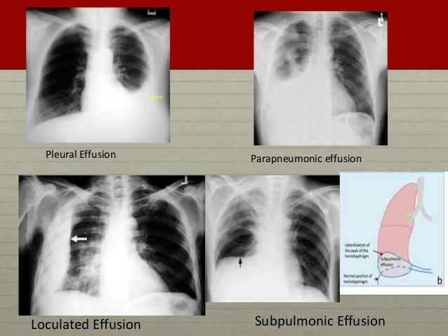
Loculated Pleural Effusion Usg - Aspiration of loculated pleural effusion - YouTube - Pleural effusion develops when more fluid enters the pleural space than is removed.
byAdmin•
0
Loculated Pleural Effusion Usg - Aspiration of loculated pleural effusion - YouTube - Pleural effusion develops when more fluid enters the pleural space than is removed.. Symptoms and causes pleural effusion prophylaxis pleural effusion. e intrinsic characteristics of an effusion and its. Pleural effusions can loculate as a result of adhesions. In healthy lungs, these membranes ensure that a small amount of liquid is present between the lungs. Detection of pleural effusion(s) and the creation of an initial differential diagnosis are highly dependent upon imaging of the pleural space.
The pleura is a thin membrane that lines the inside of the chest wall and covers the lungs. Pleura inflammation, causing sharp pain with breathing; A loculated pleural effusion are most often caused by an exudative (inflammatory) effusion. Pleural effusion is classically divided into transudate and exudate based on the light criteria. The pleura are thin membranes that line the lungs and the inside of the chest cavity and act to lubricate and facilitate breathing.
Loculated Pleural Effusion : State Of The Art Radiological ... from image.slidesharecdn.com Pleural effusion develops when more fluid enters the pleural space than is removed. e intrinsic characteristics of an effusion and its. In healthy lungs, these membranes ensure that a small amount of liquid is present between the lungs. Commonly from congestive heart failure or malignancy. Treatment depends on the cause. Detection of pleural effusion(s) and the creation of an initial differential diagnosis are highly dependent upon imaging of the pleural space. More than one half of these massive pleural effusions are caused by malignancy; A definitive diagnosis of loculated pleural effusion is best established by ultrasound.
Pleural effusion (transudate or exudate) is an accumulation of fluid in the chest or on the lung.
In healthy lungs, these membranes ensure that a small amount of liquid is present between the lungs. Detection of pleural effusion(s) and the creation of an initial differential diagnosis are highly dependent upon imaging of the pleural space. Excess fluid in the pleural space; The pleural fluid may loculate between the visceral and parietal pleura (when there is partial fusion of the pleural layers) or within. Learn about pleural effusion including causes of pleural effusion. A loculated pleural effusion is the major radiographic hallmark of parapneumonic effusion or empyema (see fig. It has many causes (pneumonia, heart failure, blood clots, trauma. Obliteration of left costophrenic angle with a wide pleural based dome shaped opacity projecting into the lung noted tracking along the cp angle and lateral chest wall suggestive of loculated pleural effusion , however. Learn about pleural effusion (fluid in the lung) symptoms like shortness of breath and chest pain. Most commonly caused by a viral infection. Learn vocabulary, terms and more with flashcards, games and other study tools. The pleura are thin membranes that line the lungs and the inside of the chest cavity and act to lubricate and facilitate breathing. Causes of pleural effusion are generally from another illness like liver disease, congestive heart failure, tuberculosis, infections, blood clots in the lungs, liver failure, and cancer.
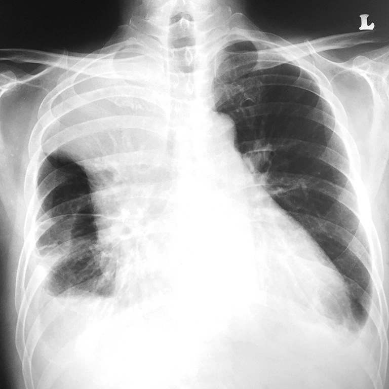
Computed tomography scan of the chest demonstrates loculated pleural effusion in the left major fissure (arrow) in a patient after coronary bypass. Pleural anatomy physiology pathogenesis of pleural effusion clinical features causes investigations treatment diagnostic approach algorithm case ultrasonography thorax this helps in detecting even the small amount of fluid. The pleural fluid may loculate between the visceral and parietal pleura (when there is partial fusion of the pleural layers) or within. Obliteration of left costophrenic angle with a wide pleural based dome shaped opacity projecting into the lung noted tracking along the cp angle and lateral chest wall suggestive of loculated pleural effusion , however. Pleural effusion occurs when too much fluid collects in the pleural space (the space between the two layers of the pleura).
Loculated Pleural Effusion Ct Scan / Pleural Effusion Pptx ... from media-us.amboss.com Accompanying adhesions can be identified. The pleura is a thin membrane that lines the inside of the chest wall and covers the lungs. e intrinsic characteristics of an effusion and its. Pleural effusions can loculate as a result of adhesions. Symptoms and causes pleural effusion prophylaxis pleural effusion. Pleura inflammation, causing sharp pain with breathing; There is normally a tiny amount of fluid between the two layers of pleura. It has many causes (pneumonia, heart failure, blood clots, trauma.
Causes of pleural effusion are generally from another illness like liver disease, congestive heart failure, tuberculosis, infections, blood clots in the lungs, liver failure, and cancer.
Diffuse nodules and opacification in right lung with compressive atelectasis. Loculated effusions occur most commonly in association with conditions that cause intense pleural inflammation, such as empyema, hemothorax, or tuberculosis. Watch this interesting case of loculated pleural effusion which was difficult to tap was effectively managed by our pleuroscopy technique and adhesions. Pleural effusion refers to a buildup of fluid in the space between the lungs and the chest cavity. Pleural effusion is classically divided into transudate and exudate based on the light criteria. Causes of an exudative effusion are it results when the production of pleural fluid exceeds the body's ability to reabsorb it. There is normally a tiny amount of fluid between the two layers of pleura. Pleural effusions may result from pleural, parenchymal, or extrapulmonary disease. Benefits of chest ct for effusion. Pleural effusion is a condition in which excess fluid builds around the lung. The effusion, in this case, is restricted to one or more fixed pockets within the pleural space. Meaning of pleural effusion medical term. Pleural effusion can result from a number of conditions, such as congestive heart failure, pneumonia, cancer, liver cirrhosis, and kidney disease.
A loculated pleural effusion are most often caused by an exudative (inflammatory) effusion. Most commonly caused by a viral infection. Approximately 1 million people develop this abnormality each year in the united states. Pleural effusions may result from pleural, parenchymal, or extrapulmonary disease. Benefits of chest ct for effusion.
Loculated transudative pleural effusion masquerading as ... from cdn.amegroups.cn Usg is helpful in cases of loculated pe for confirmation of the diagnosis. Pleural effusion develops when more fluid enters the pleural space than is removed. Other causes are complicated parapneumonic effusion. Symptoms and causes pleural effusion prophylaxis pleural effusion. Treatment depends on the cause. Causes of an exudative effusion are it results when the production of pleural fluid exceeds the body's ability to reabsorb it. Pleural effusions can loculate as a result of adhesions. Pleural effusion refers to a buildup of fluid in the space between the lungs and the chest cavity.
Usg is helpful in cases of loculated pe for confirmation of the diagnosis.
The pleura is a thin membrane that lines the surface of your lungs and the inside of your chest wall. Meaning of pleural effusion medical term. Pleural effusions occur as a result of increased fluid formation and/or reduced fluid resorption. It has many causes (pneumonia, heart failure, blood clots, trauma. The pleura are thin membranes that line the lungs and the inside of the chest cavity and act to lubricate and facilitate breathing. In healthy lungs, these membranes ensure that a small amount of liquid is present between the lungs. Accompanying adhesions can be identified. Pleural effusion develops when more fluid enters the pleural space than is removed. Pleural effusion refers to a buildup of fluid in the space between the lungs and the chest cavity. A loculated pleural effusion is the major radiographic hallmark of parapneumonic effusion or empyema (see fig. Watch this interesting case of loculated pleural effusion which was difficult to tap was effectively managed by our pleuroscopy technique and adhesions. Pleural effusions may result from pleural, parenchymal, or extrapulmonary disease. Pleural effusions are a common medical problem with more than 50 recognised causes including disease local to the pleura or underlying lung, systemic conditions, organ dysfunction and drugs.1.
A loculated pleural effusion is the major radiographic hallmark of parapneumonic effusion or empyema (see fig loculated pleural effusion. e intrinsic characteristics of an effusion and its.